"Be Breast Aware."
AWARENESS IS POWER!
70-80% of cancer cases in Kenya are diagnosed at advanced stages.According to Globacon 2020 WHO’s Agency for research on cancer, Breast cancer is one of the most commonly diagnosed cancers in Kenya, with an annual incidence of 6,000 new cases and 2,500 cancer-related deaths.
Late-stage diagnosis is one of the greatest impediments in making significant progress in combating cancer. The World Health Organization (WHO) champions awareness, early detection and screening such as screening mammograms & pap smears as the cornerstone of breast & cervical cancer control respectively.
Awareness, Early diagnosis and Affordable treatment could greatly reduce the burden of cancer and improve treatment outcomes.
Early Detection, Saves Lives.

Awareness Equals +Change
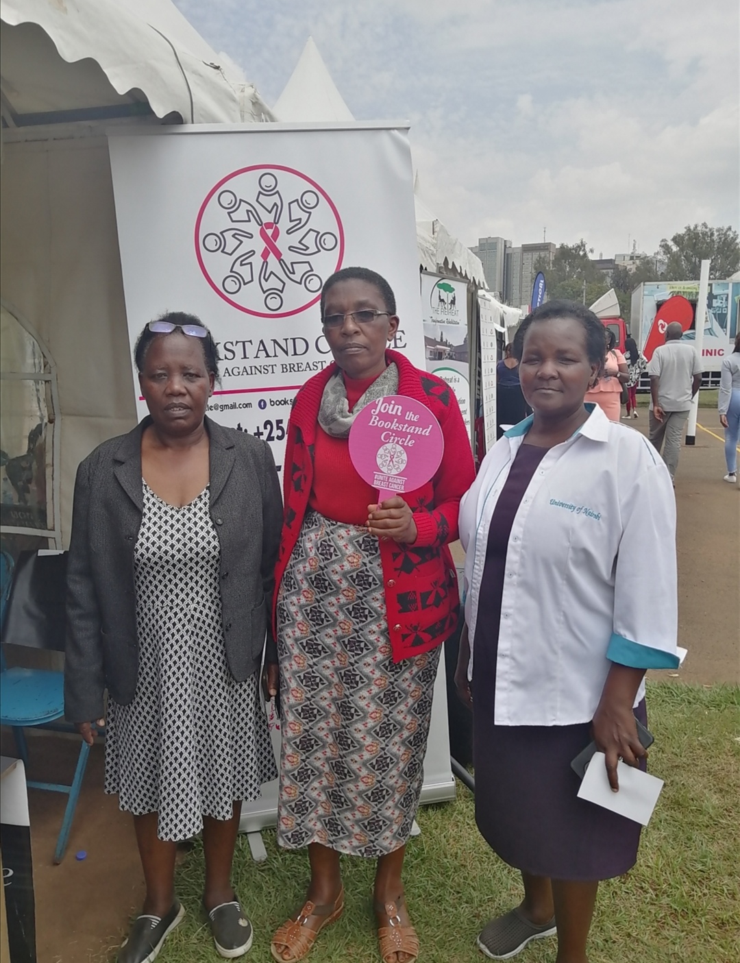
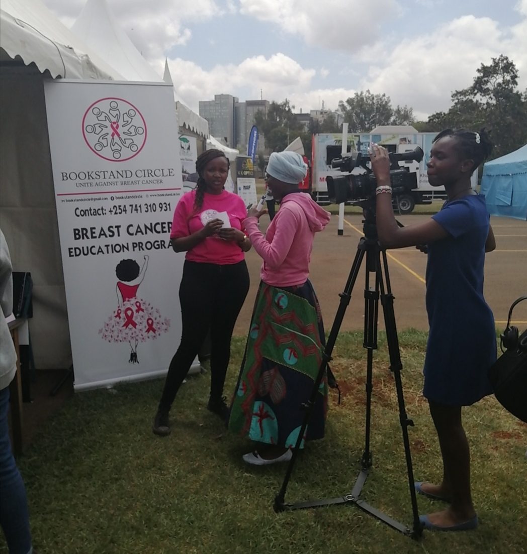
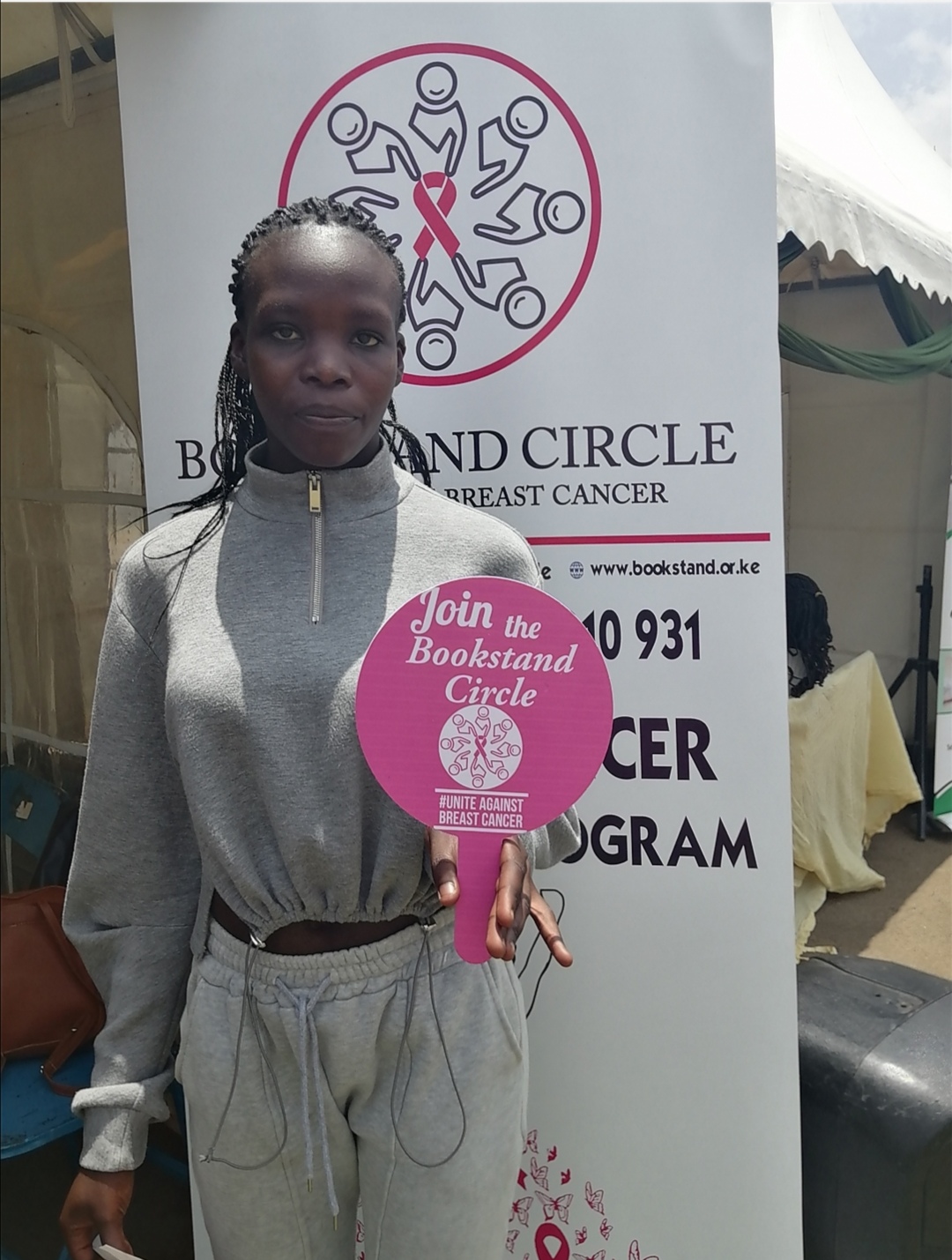
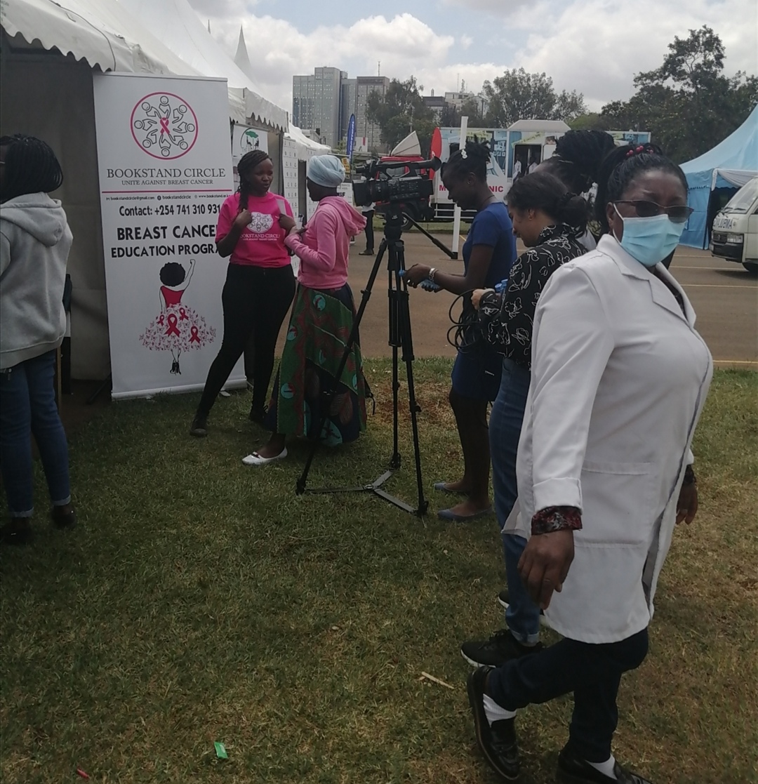
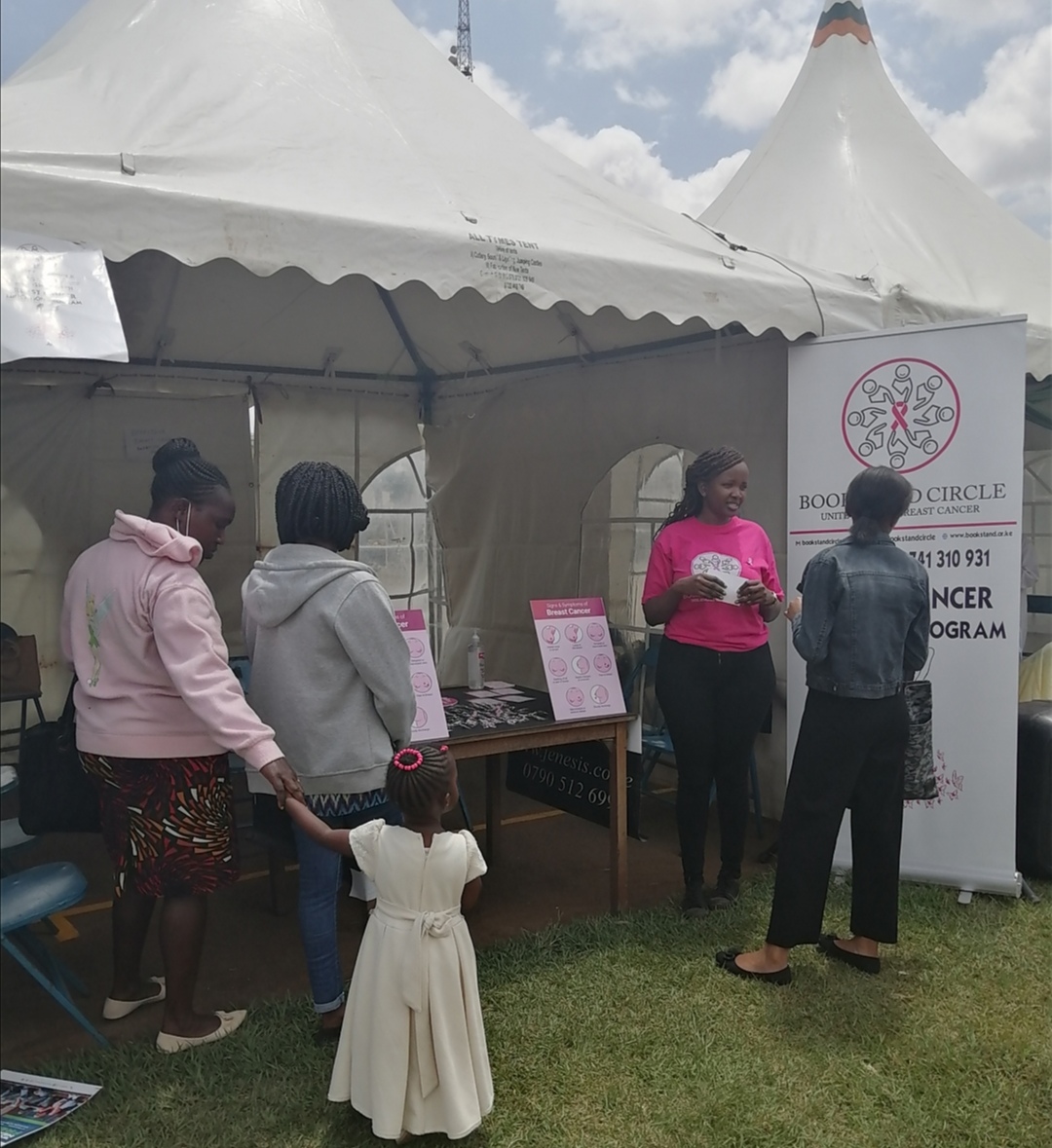
Champions of Change aim to advance breast and cervical cancer awareness through youth engagement and awareness camps targeting underserved/low income women. Join the movement . Call/email us Today
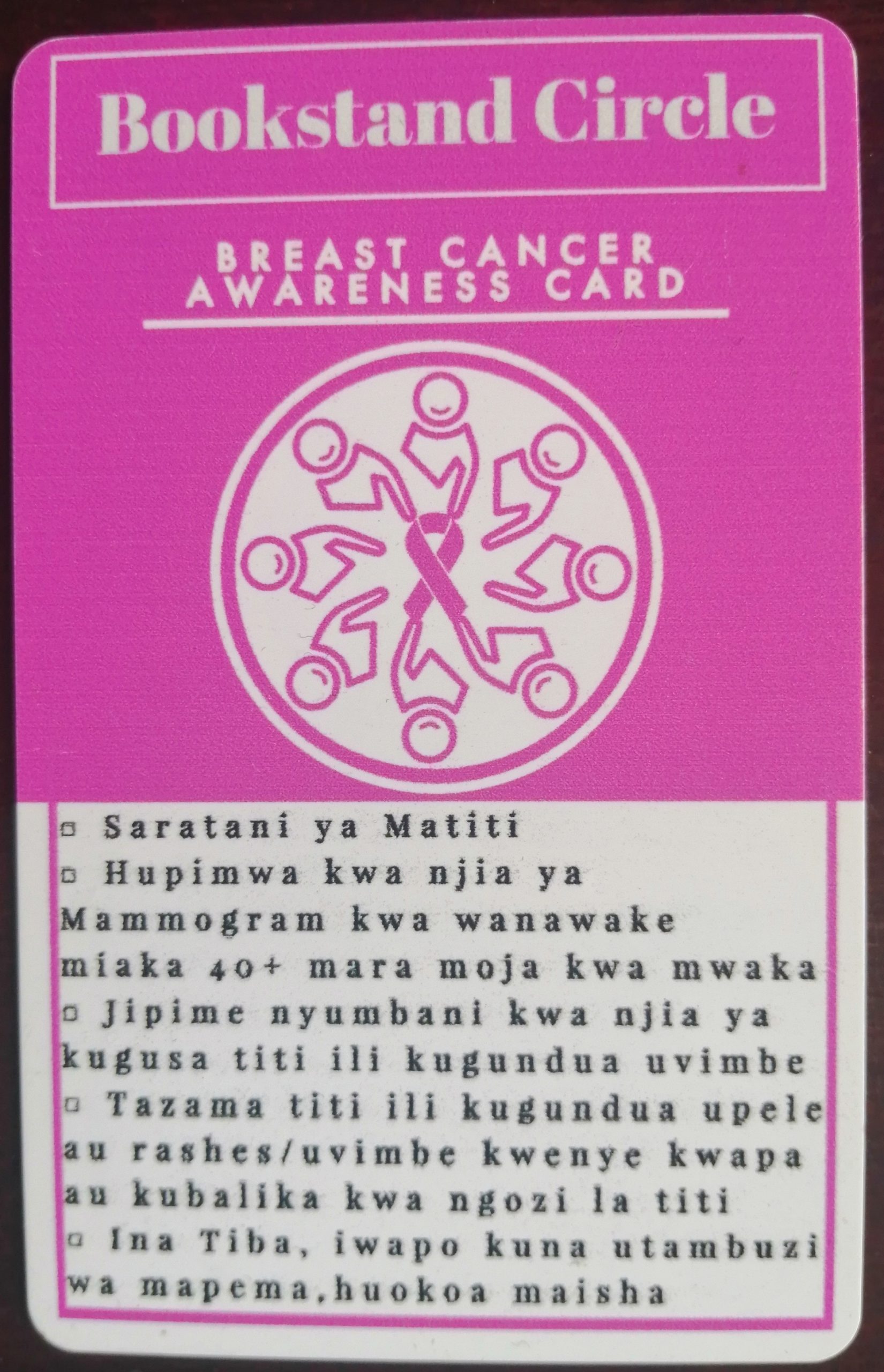
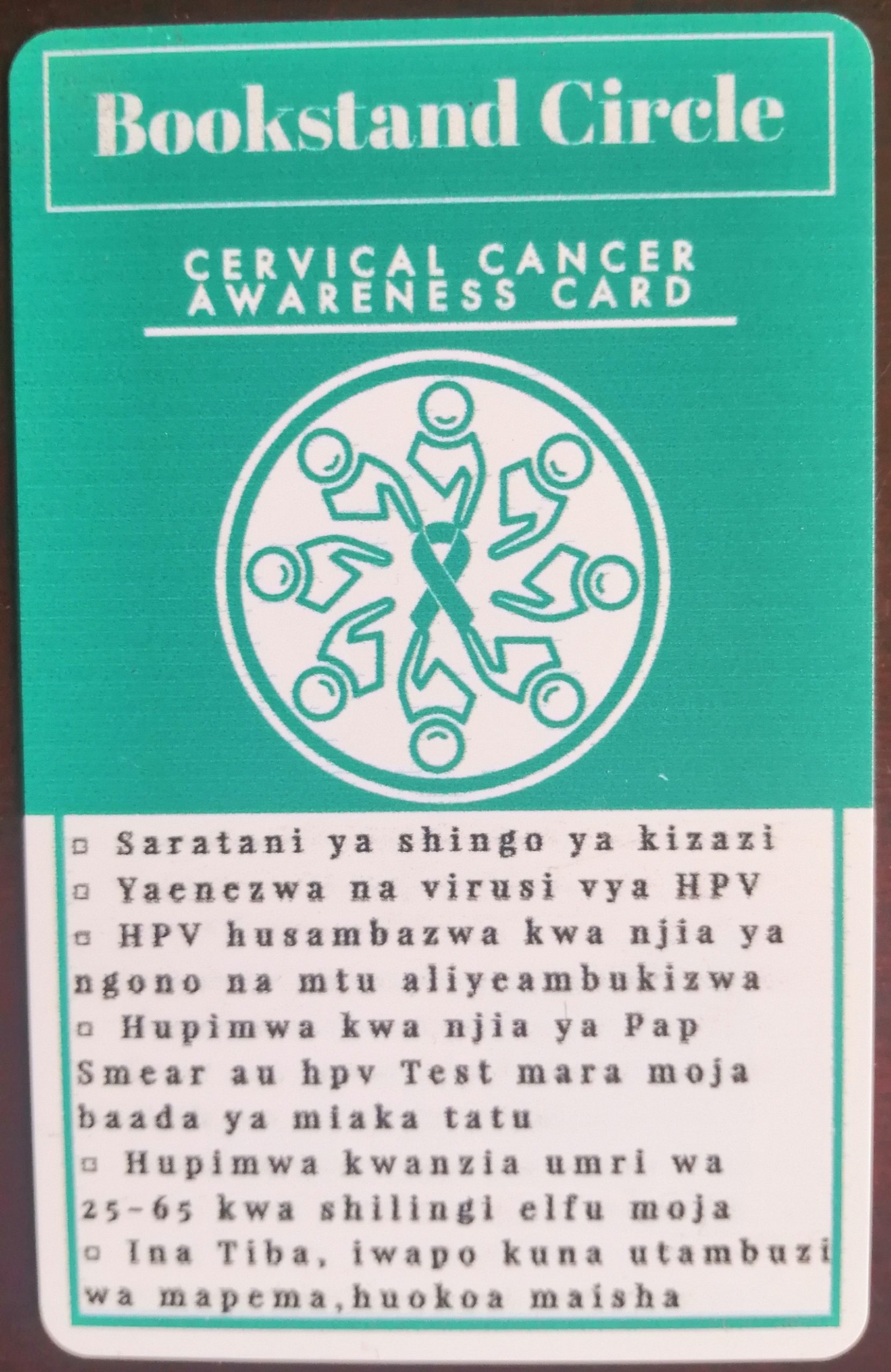
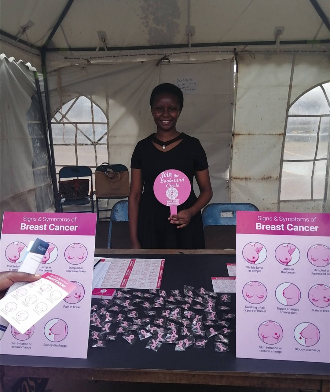
Cancer Awareness Cards
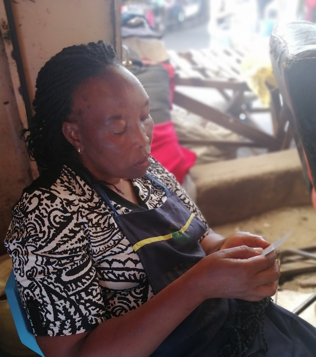
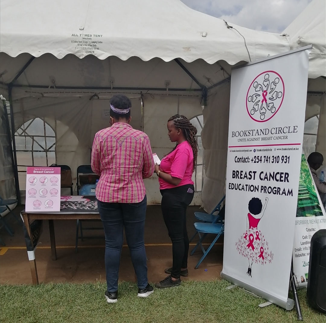
The Bookstand cancer awareness cards are durable pvc cards that educate the public on the causes, early detection symptoms, prevention and screening of breast and cervical cancer. Partner/volunteer with us as we soldier on to raise awareness and have a positive impact within the undeserved demographic.


AWARENESS CAMPAIGN IN PARTNERSHIP WITH THE UNIVERSITY OF NAIROBI HEALTH SERVICES
Awareness is the greatest agent for change. Early Detection is your Best Protection.


The Pink Beyond October Campaign
As amazing as breast cancer awareness month is for generating support for the cause during October, the reality is that breast cancer is a 365 day concern. At Bookstand, we are always thinking of new ways to empower young people and women to learn about breast health and breast awareness throughout the year. We hope you will join us and support the breast cancer movement beyond October! Donate in support of the beyond October awareness program.
What is Oncology?

Oncology is the study of cancer. An oncologist is a doctor who treats cancer and provides medical care for a person diagnosed with cancer. An oncologist may also be called a cancer specialist.
The field of oncology has 3 major areas based on treatments: medical oncology, radiation oncology, and surgical oncology.
- Medical oncologists treat cancer using medication, including chemotherapy, immunotherapy, and targeted therapy.
- Radiation oncologists treat cancer using radiation therapy, which is the use of high-energy x-rays or other particles to destroy cancer cells.
- Surgical oncologists treat cancer using surgery, including removing the tumor and nearby tissue during a operation. This type of surgeon can also perform certain types of biopsies to help diagnose cancer.
An oncologist manages a patient’s care throughout the course of the disease. This starts with the diagnosis. Their role includes:
Recommending tests to determine whether a person has cancer
Explaining a cancer diagnosis, including the type and stage of the cancer
Talking about all treatment options and your treatment choice
Delivering quality and compassionate care
Helping you manage symptoms and side effects of cancer and its treatment
Radiologists are doctors who read mammograms and other types of imaging tests, they check for abnormalities, calcifications, masses, asymmetries, breast density and distortions.
Frequently Asked Questions.
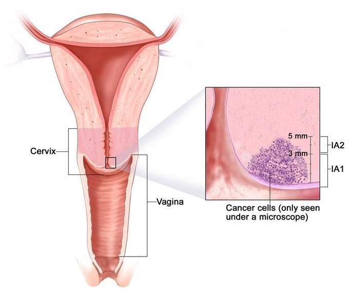
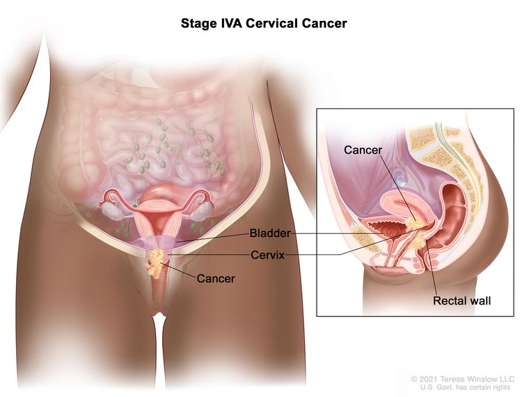
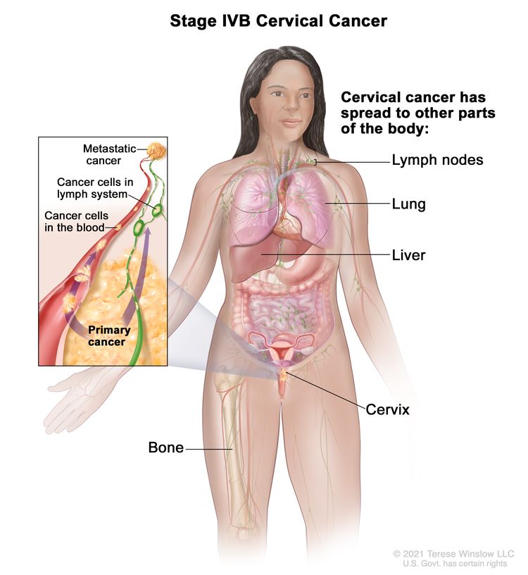
Cervical cancer begins when healthy cells on the surface of the cervix change or become infected with human papillomavirus (HPV) and grow out of control, forming a mass called a tumor. Long-term infection of HPV on the cervix can result in cancer, leading to a mass or tumor on the cervix.
EARLY SYMPTOMS:
- Unusual bleeding from the vagina like bleeding when you’re not on your period, after menopause or after sex
- Unpleasant smelling vaginal discharge
- Pain and discomfort after sex
Unexplained, persistent pelvic and/or back pain
It may feel like a sharp pain or pressure, located anywhere around the lower abdomen, below the belly button.
ADVANCED SYMPTOMS:
- Leg pain that feels like a persistent sharp or dull ache
- Swelling in the legs
- Weight loss
- Fatigue
- Back pain
- Leakage of urine or feces from the vagina
- Bone fractures
- Difficulty urinating and having a bowel movement
- Blood in the urine
The HPV test and the Pap smear test can help detect cervical cancer or find it early.
The HPV test looks for the virus (human papillomavirus) that can cause cell changes on the cervix. The Pap test (or Pap smear) looks for precancers, cell changes on the cervix that might become cervical cancer if they are not treated appropriately.
- Begin screening at age 25 to 65.
Doctors generally recommend repeating Pap testing every three years for women ages 21 to 65.
Women age 30 and older can consider Pap testing every five years if the procedure is combined with testing for HPV. Or they might consider HPV testing instead of the Pap test.
- There are three types of cancer treatment: Surgery, Radiotherapy and Chemotherapy, additionally immunotherapy and targeted therapy
- Cervical cancer is highly preventable and highly curable if caught early. Nearly all cervical cancers could be prevented by HPV vaccination routine cervical cancer screening, and appropriate follow-up treatment when needed.

01
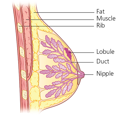
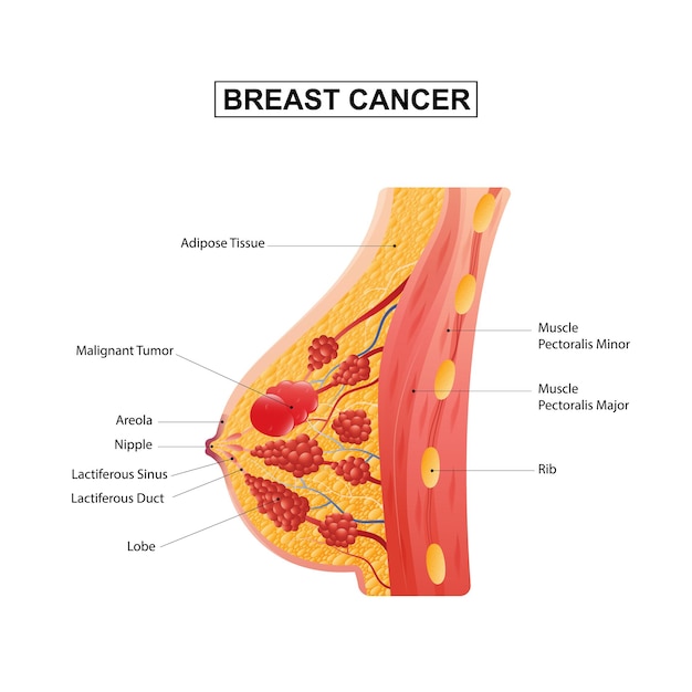
Breast cancer forms in either the lobules or the ducts of the breast. Lobules are the glands that produce milk, and ducts are the pathways that bring the milk from the glands to the nipple. Cancer can also occur in the fatty tissue or the fibrous connective tissue within your breast. The cancerous cells can spread outside the breast through blood vessels and lymph vessels. When breast cancer spreads to other parts of the body, it is said to have metastasized.

Frequently Asked Questions.

Breast cancer occurs when malignant (cancerous) tumors develop in the breast. Tumors may be either benign (non-cancerous) or malignant (cancerous). Benign tumors usually grow in one place and do not spread. Malignant tumors develop in one area of the body, then spread to other organs effectively damaging them.
- Lumps In The Breast Or Underarm
- Bloody Nipple Discharge
- Nipple Retraction
- Dimpling Skin/change in the skin texture
- Itching, Flaking Or Crusting Of The Nipple
- New pain in one area of the breast that does not go away
- There are three types of cancer treatment: Surgery, Radiotherapy and Chemotherapy, additionally hormone therapy and targeted therapy
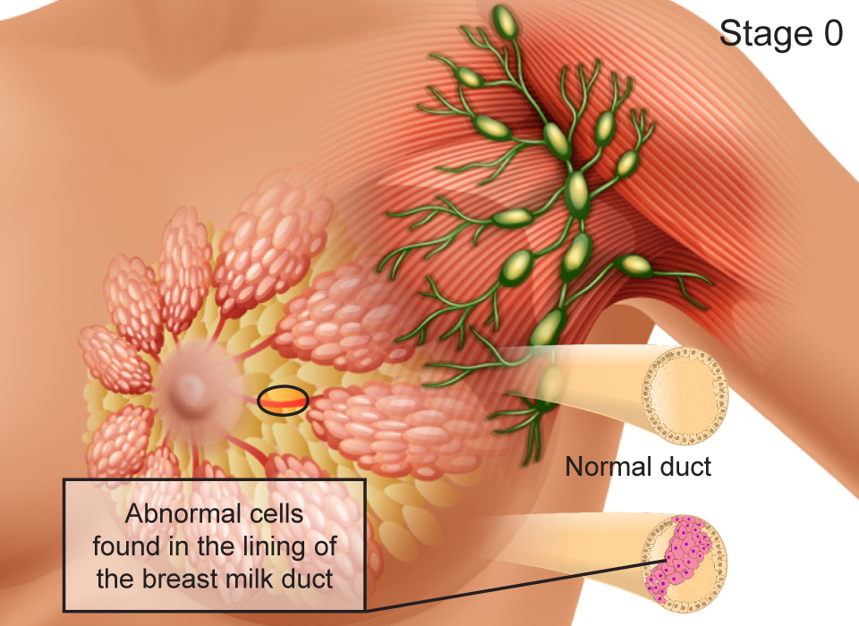
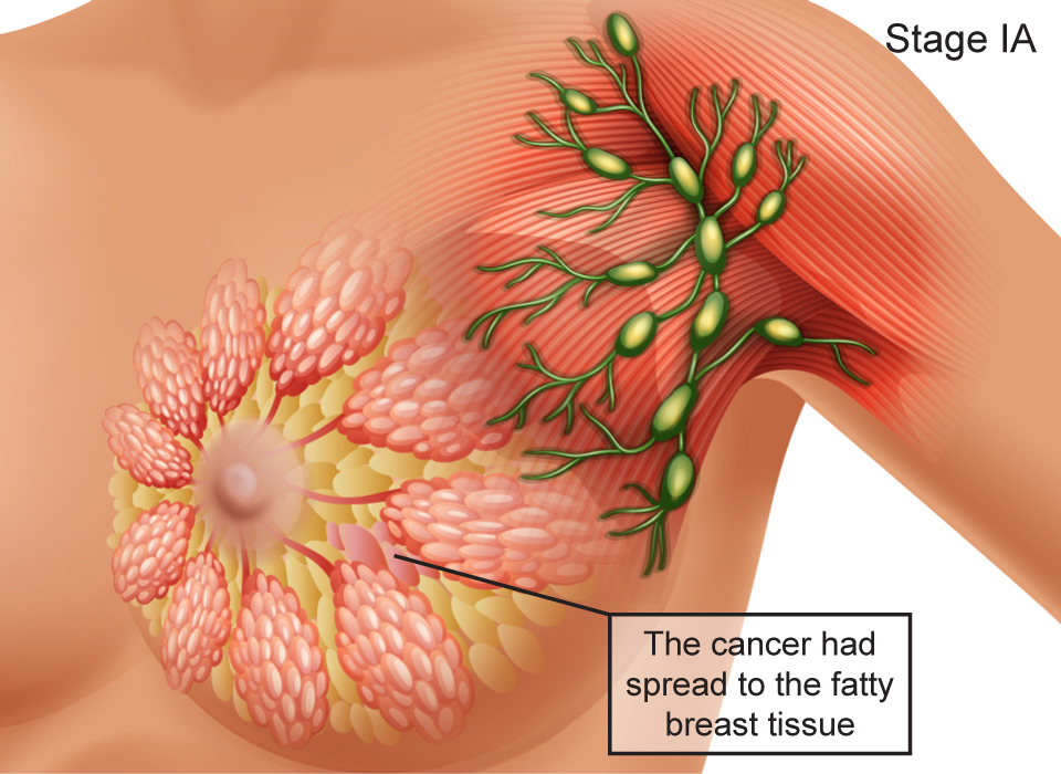
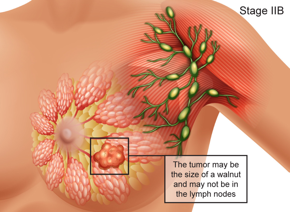
- Stage 0: is very early cancer that is highly treatable, but if it’s left untreated or undetected, it can spread into the surrounding breast tissue.
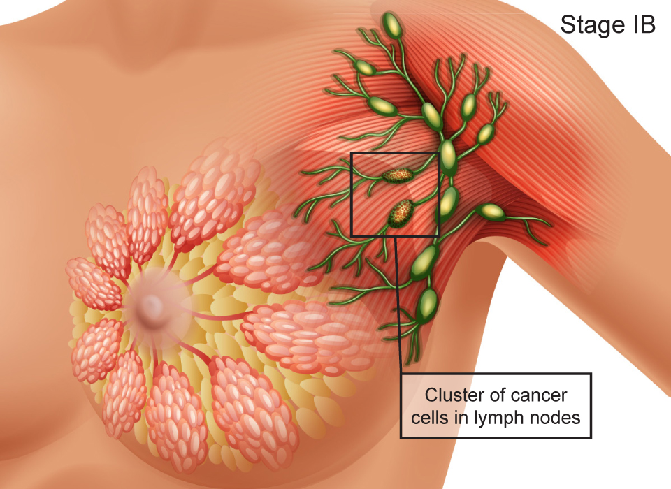
- Stage 1: Though considered “non-invasive,” it does require treatment, typically surgery or radiation, or a combination of the two.
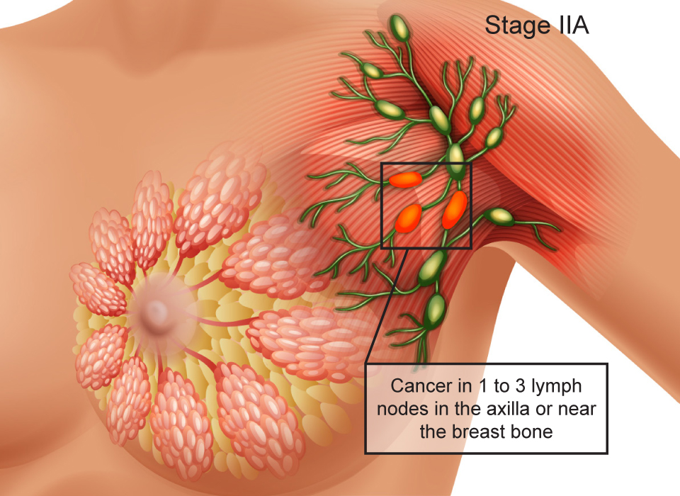
- Stage 2: Chemotherapy is usually done first, followed by surgery and radiation therapy.
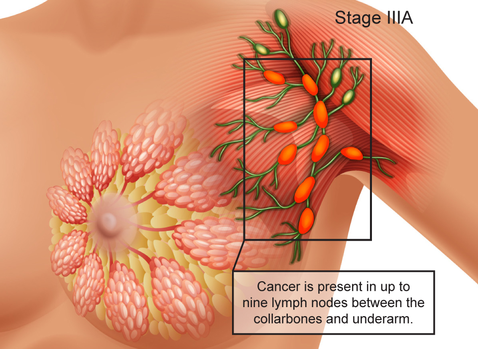
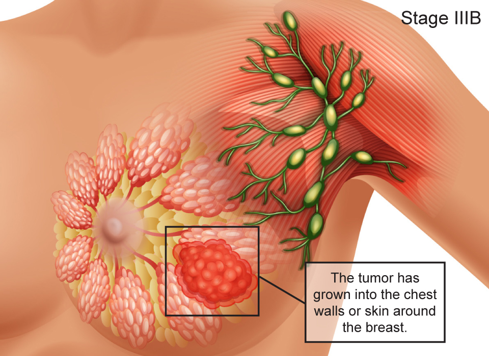
- Stage 3: Treatment options vary widely and may consist of mastectomy (a surgical operation to remove the breast) and chemotherapy (shrinks the cancerous cells) and radiation (kills cancerous cells).
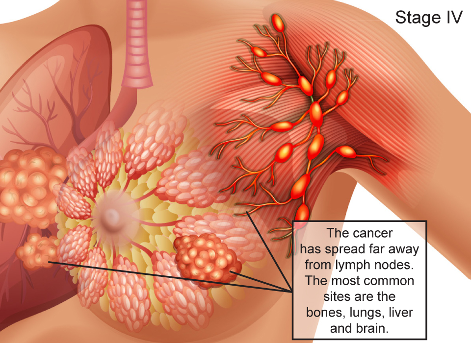
- Stage 4: Seek out oncology specialists who specialize in Stage 4 breast cancer. Discuss with your treatment team what clinical trials may be available for your clinical situation. You can consider trying therapies that are still in the experimental phase.
- Find Out Your Family History
- Breast self exam once a month
- Keep healthy weight
- Limit or avoid alcohol
- Be physically active
- If possible Breastfeed
- Visit a hospital for mammograms
- Eat healthy
- Most breast lumps are not cancerous
- Most breast lumps are fluid filled cysts or fibroadenomas which are benign
- However you should always see a breast cancer treatment doctor if a lump develops as the breast lump may be cancerous
BREAST CANCER STAGES
What is a Clinical Breast Exam?
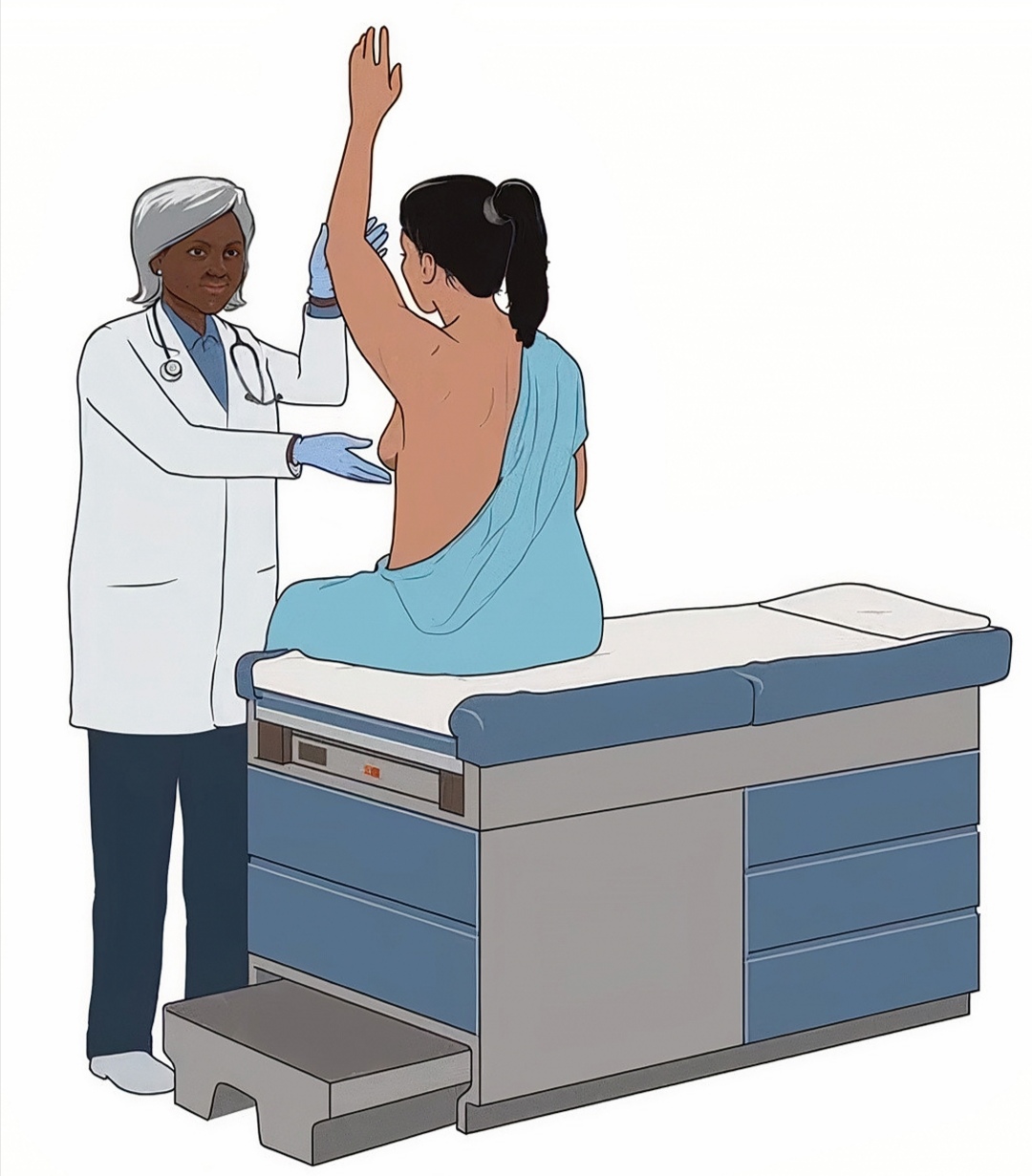
A CBE is a physical exam of the breast performed by a health care provider to check for lumps or other changes.
A CBE is a key step in the diagnosis and surveillance of a number of benign (non-cancerous) and malignant (cancerous) breast diseases. The goal of the breast examination is to determine if the breasts are normal or abnormal. It also provides the opportunity to educate women on self breast examination and early detection symptoms of breast cancer.
Frequently Asked Questions.
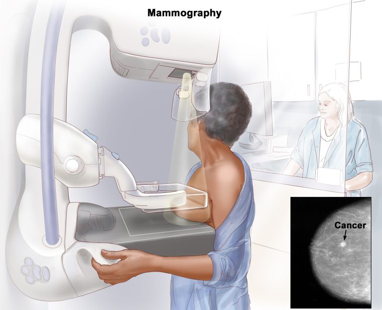
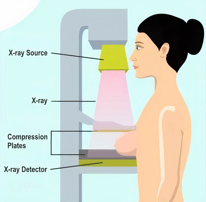
A mammogram is a special type of X-ray that captures images of the breast tissue to help detect tumors. It is one of the most effective ways to detect and screen for Breast Cancer in the initial stages
Mammography is chiefly recommended for every woman above the age of 40, in every 1 or 2 years. But in case, you have a personal or family history of breast cancer, the doctor may recommend you to start breast screenings earlier in life, and have them more often, or use additional diagnostic tools.
- Depending upon the diagnosis, mammography is of two types:
- Screening Mammography: It is the procedure that is done as a routine test to check for any cancer or changes in the breast.
- Diagnostic Mammography:A procedure which is conducted if you have a lump or any other symptom of breast cancer.
- While the process for both is same, diagnostic mammography is more extensive than screening mammography and may require more images than the screening.
- The person undergoing a mammography need to follow certain guidelines on the day of the procedure to avoid glitches in the report:
- The person must not apply any deodorant, body powder, ointments, creams or perfumes on the breasts or underarms as they may create unnecessary white spots on the image.
- The person undergoing the mammography is suggested to use the same facility every year to make comparison of the results easier. In the case, you are using a facility or diagnostic centre for the first time, do ensure that you carry your previous mammograms.
- Make sure to describe the treating doctor or the technician about any breast symptoms or problems prior to the procedure.
- Try not to conduct mammography the week before you get your period or during the menstrual cycle as your breasts might be tender or swollen at the time.
- Do not forget to mention the treating doctor or radiologist in case you are pregnant or breast feeding, as it is not good to get exposed to X-ray during this time. But if necessary or depending upon the severity of the condition, the doctor can recommend other screening methods, such as an ultrasound.
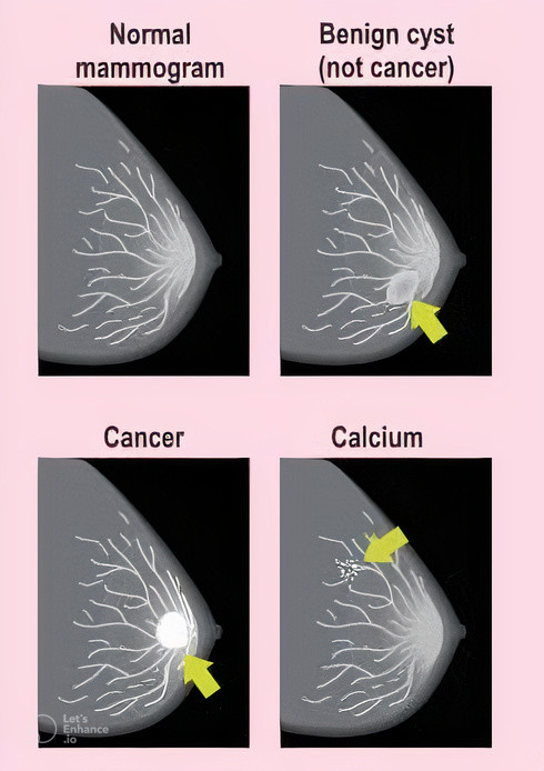
Mammography, uses low-energy X-rays to examine the human breast for early detection of breast cancer typically through detection of characteristic masses or microcalcifications. It is recommended alongside regular clinical exams and monthly breast self-examinations.
- The breasts are placed or fitted on a resting plate, and a compression device is used to push the breast down to flatten the tissue to get a clearer picture of the breast.
- The person may feel some amount of discomfort but it is usually temporary and does not cause any damage to the breast tissue.
- Typically, the technician takes two views of each breast. Once the films are developed, these radiographs are checked by the technicians for clinical accuracy before the person leaves.
- Once the procedure is conducted, the person will usually get the films within a week.
- The films are then examined by radiologists who has specialized training in the interpretation of breast images.
- Apart from detection of cancer, a mammography can help find calcifications, or calcium deposits, in the breasts. It can also find cysts within the breast tissue which may come and go normally during some people’s menstrual cycles and also presence of any cancerous or noncancerous lumps.
- If the mammography is normal, continue to do the process every year as a routine check-up.
- If the results are abnormal, the doctor may suggest the patient to go for additional mammograms, tests, exams or other imaging techniques such as MRI or Ultrasound. The doctor may also refer the patient to a specialist or a surgeon in case the procedure detects cancer and the person needs a surgery.
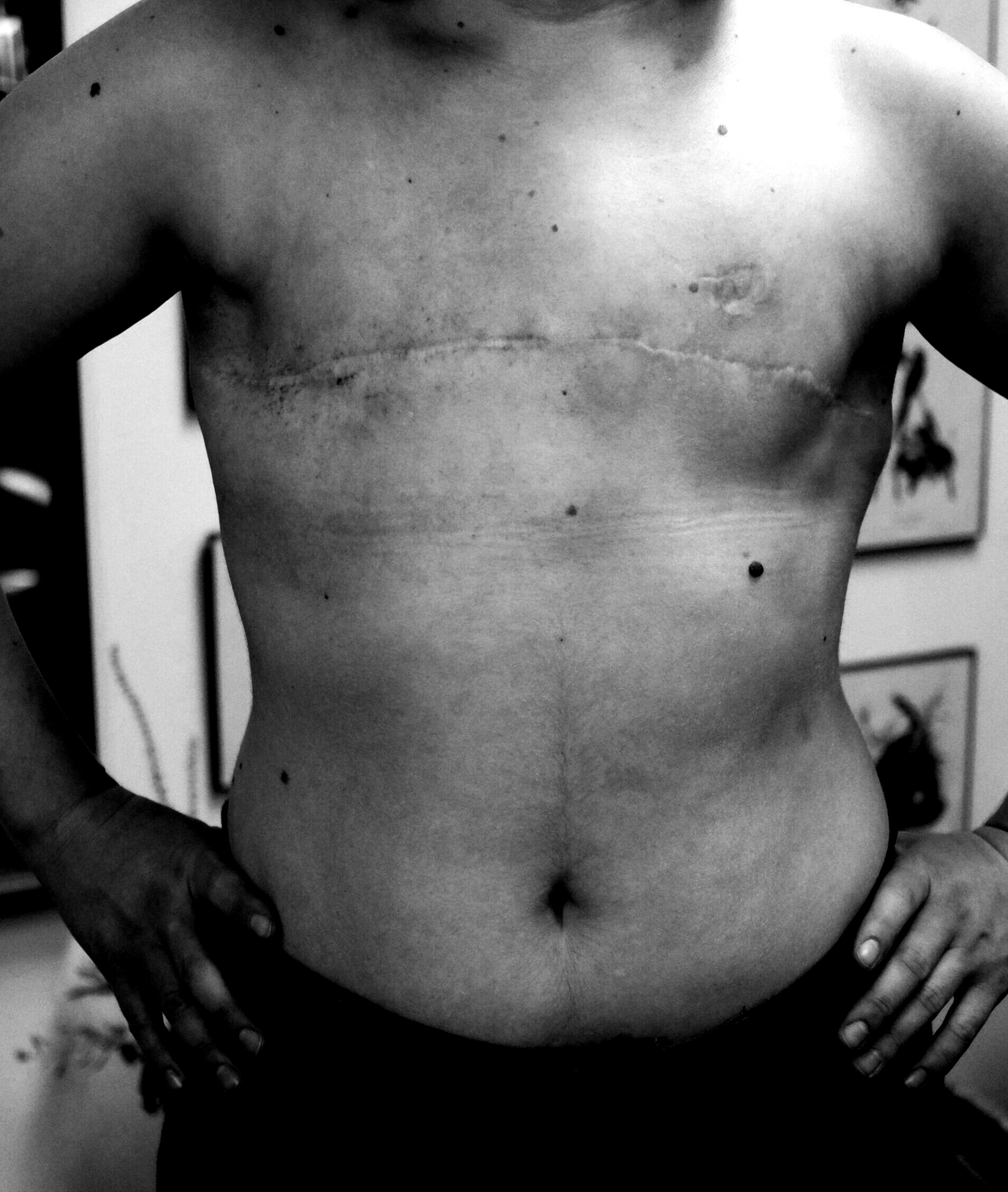
Mastectomy
A mastectomy is a major surgey used to treat breast cancer by removing part or the entire breast through surgery. In some cases, women believed to be at high risk of breast cancer have the operation as a preventive measure, however it is best to consult with your oncologist on the best treatment for your diagnosis.

02
When it comes to the early symptoms of breast cancer, it’s not all about lumps. Breast cancer can make itself known via other lesser-known symptoms. That’s why it’s important NOT to dismiss any odd new developments on your nipples or breasts, even if you don’t feel any lumps; It is important to become familiar with your own body and be able to recognize any changes.
Regular self examination of breasts is an effective way to detect signs of cancer. The best time to practice self-examination is 3-5 days after the menstrual cycle. During the menopause phase, self-examination can be done on the same day of the month.
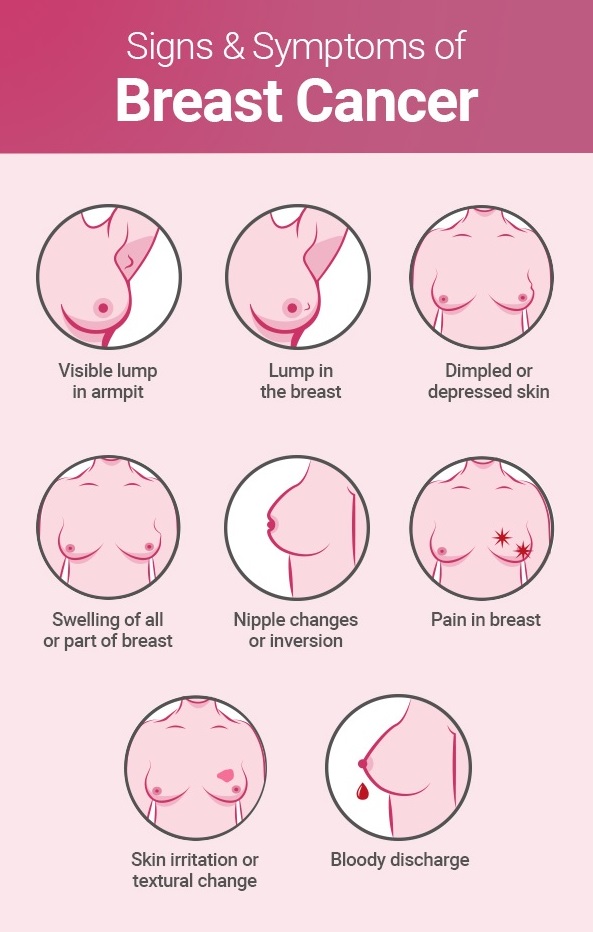
Early Signs & Symptoms of Breast Cancer
Early signs of breast cancer show up when the cancer is new, and may not have developed to a more advanced stage that requires more intensive treatment.
It’s important to detect the symptoms early which significantly increases your chances of successfully beating it. Breast cancer that’s found and treated when it’s still at stage 1A (a cancer smaller than a peanut that hasn’t spread anywhere) has a survival rate of 100%, according to the National Breast Cancer Foundation.
How to Perform a Self Breast Exam

Stand in front of a mirror with your shoulders straight and your hands on your hips. Examine if the breasts and nipples are their usual size, shape and color in the mirror. Also examine if there is any swelling, redness and rashes on the breast skin. Also look for any change in the position of the nipples.
Stretch arms over the head and examine breasts in the mirror once again for the same signs as mentioned above. This process can also be done in the shower. If you observe any of the changes listed above, bring it to your doctor’s attention immediately.
Check for any fluid discharge from the nipples (watery, milky, yellow fluid or blood). Healthy breasts should not secrete fluid (unless you are lactating).
Lay on your back to examine breasts. Use the right hand to examine the left breast, and vice-versa. Keep fingers flat together and use circular or vertical motion to examine the entire breast from the collarbone to the waist and from the armpit to the cleavage.
Examine breasts while standing or sitting down, by following the method mentioned in step 4.
KENYA CANCER CARE HOSPITALS